
Fetal development week by week BabyCenter
Fetus in fetu (FIF) is a rare congenital anomaly with an incidence of 1 in 500,000 live births with male predominance. It occurs from anomalous embryogenesis in a diamniotic monochorionic twin pregnancy in which a malformed monozygotic twin lies within the body of fellow twin. The most common location is the retroperitoneum and the other being.
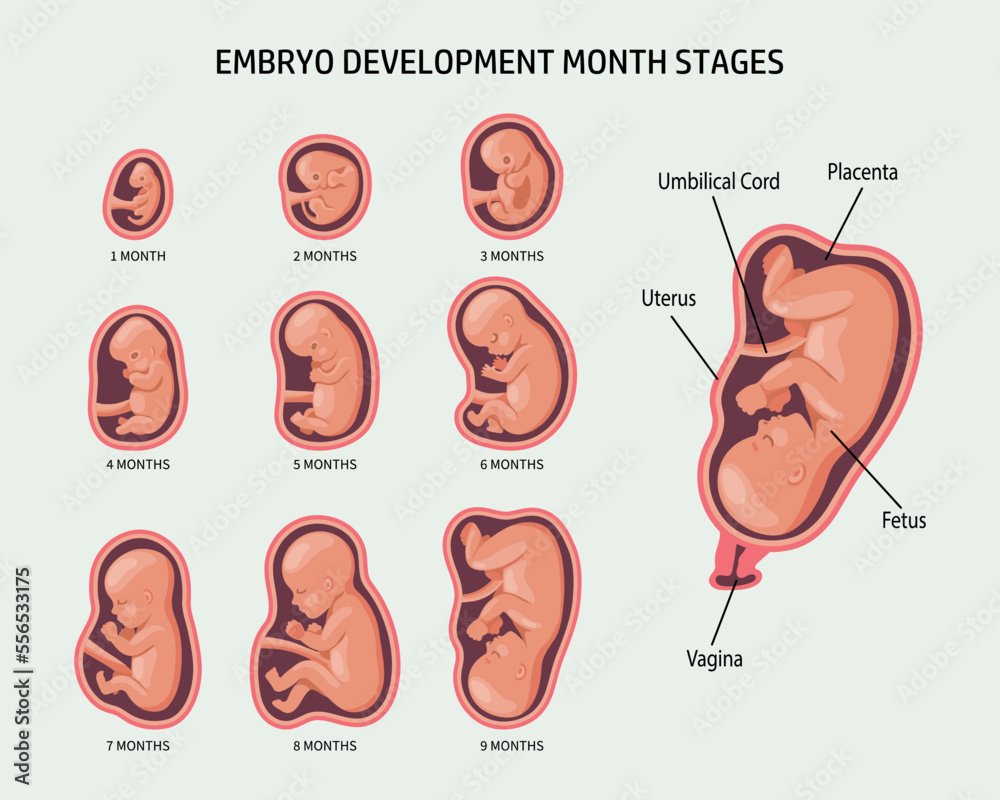
Embryo in the womb, set. Development and growth of the fetus at
Fetus in fetu is a rare condition which most often presents as a fetiform calcified mass in the abdomen of its host, fetus or newborn. We report a case of 8-month-old girl with history of abdominal distension. Ultrasonography and computed tomography scan revealed a mass in which the contents favor a fetus in fetu rather than a teratoma.

Two cases of fetus in fetu Journal of Pediatric Surgery
Introduction "Fetus in fetu" (FIF) is defined as the abnormal monozygotic twin inside the body of its "host twin." Intracranial FIFs are extremely rare. Case presentation A male premature newborn was admitted to the hospital due to a large intracranial tumor diagnosed in the 31st week of gestation. The child died before surgical treatment because of failure of the respiratory system.

Normal Anatomy of the Fetus at MR Imaging RadioGraphics
Fetus in fetu (FIF) is a rare congenital anomaly with an incidence of 1 in 500,000 live births. It was first described by Meckel, a German anatomist. FIF is a malformed parasitic twin found inside the body of its host co-twin, usually in the abdominal cavity. It is formed as a consequence of an unequal division of the totipotent inner cell mass.

Normal Anatomy of the Fetus at MR Imaging RadioGraphics
Fetus-in-fetu (FIF) is a rare entity in which one malformed vertebrate fetus is enclosed within the body of its twin. This is an extremely rare condition, and Hopkins et al. found less than 100 case reports on extensive review of the literature. An array of presentations is described in the literature although the embryo-pathogenesis and.

Normal Anatomy of the Fetus at MR Imaging RadioGraphics
1. Background. Fetus in fetu is a rare congenital entity with an incidence of 1 in 500000 births. It is a monozygotic diamniotic twin where the parasitic twin develops inside the body of a host twin [1].Most of the cases are diagnosed before 18 months of age [2].It is found most commonly in the retroperitoneum [3, 4].Atypical locations like skull, sacrum, scrotum and mouth has also been.

Placenta’s alarm clock signals when it’s time for birth to begin New
Fetus in fetu is a rare variety of parasitic twins, where the developmentally abnormal parasitic twin is completely encapsulated within the torso of the otherwise normally developed host twin. In the late eighteenth century, German anatomist Johann Friedrich Meckel was the first to described fetus in fetu, which translates to fetus within fetus.Fetus in fetu is thought to result from the.

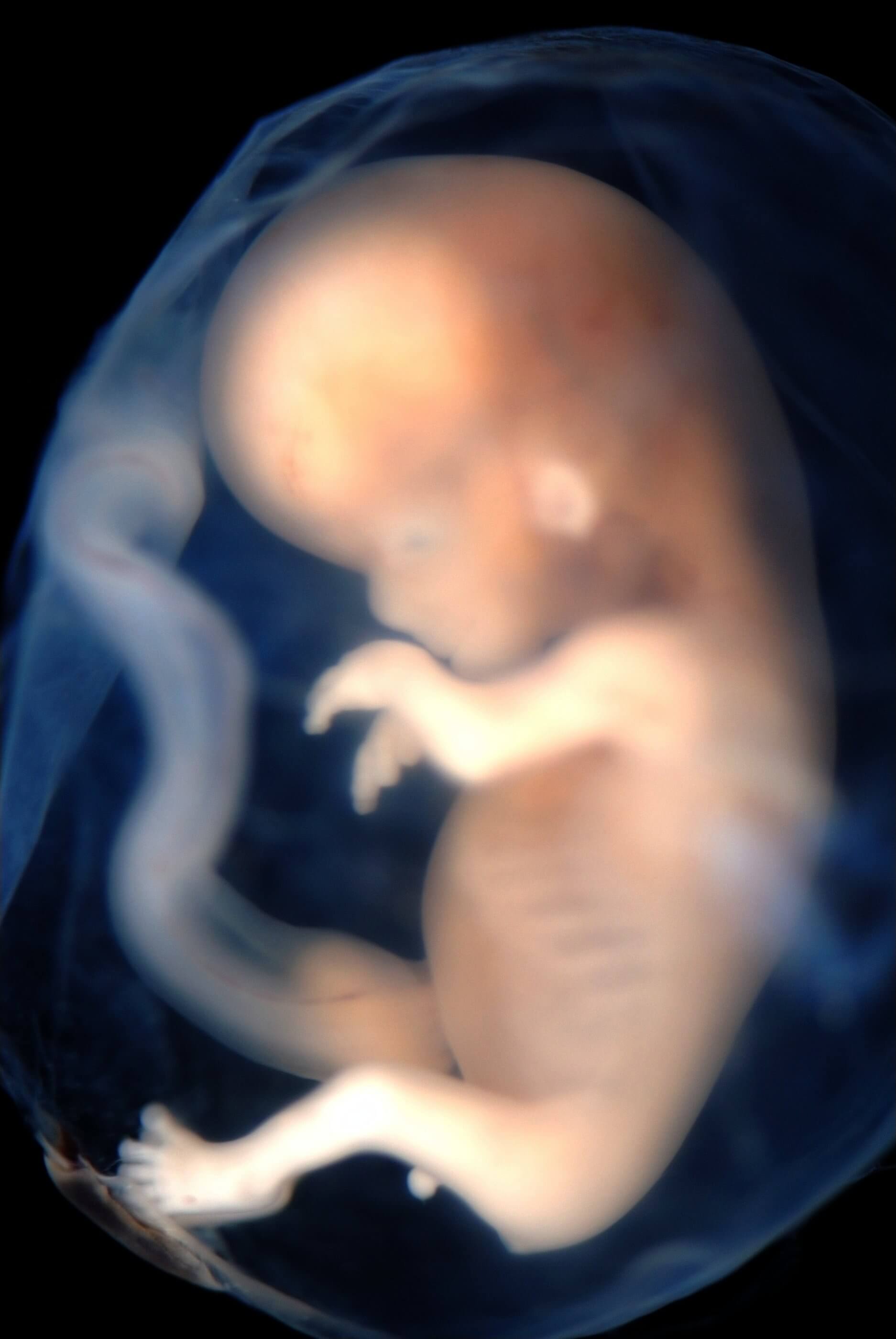
Fetus 4 Months Stock Photo Download Image Now iStock
Fetus-in-fetu is a rare entity estimated to occur in 1:500,000 deliveries, with fewer than 100 cases reported worldwide [].Generally, fetus in fetu is a single parasitic twin, but there can be multiple fetuses-in-fetu sometimes [].It is predominantly retroperitoneal in 80% of cases, while reported uncommon sites include the oral cavity, sacrococcygeal region, and scrotum [3, 4].
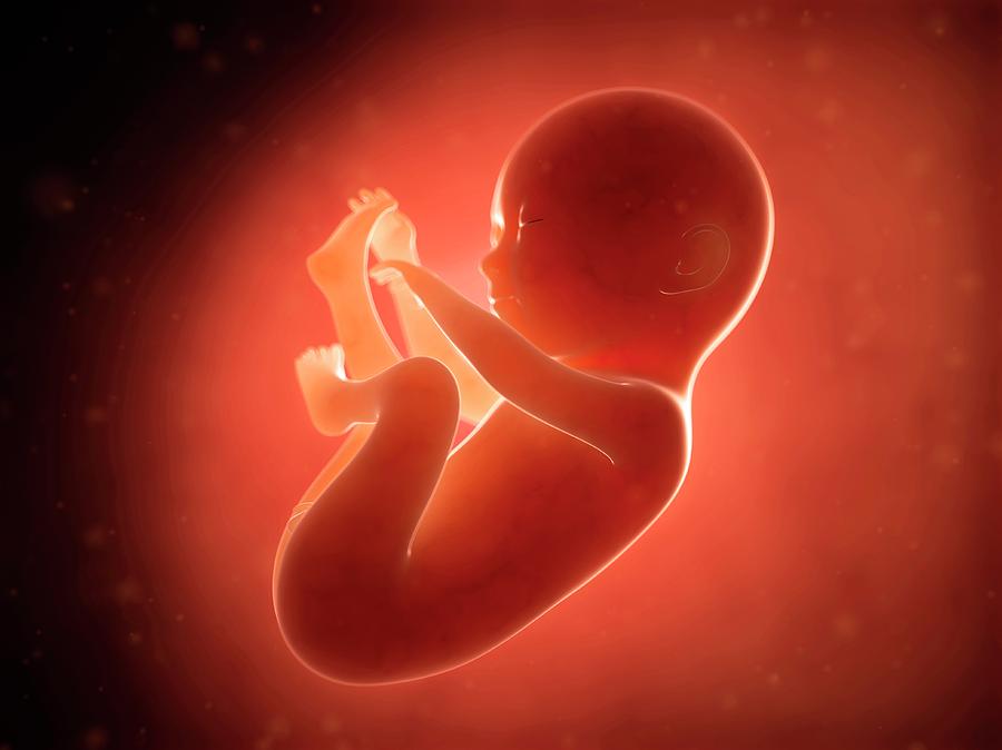
Human Fetus At 6 Months Photograph by Sebastian Kaulitzki Fine Art
Fetus-in-fetu (FIF) is an extremely rare congenital anomaly in which a vertebrate fetus-like mass is situated within the body of its fully developed host. Two main hypotheses on the pathogenesis of FIF have been described. The "included-twin" or "parasitic-twin" theory suggests that FIF arises from an anomalous monozygotic monochorionic.

Revisiting fetusinfetu Annals of Saudi Medicine
The growth of a fetus in fetu initially parallels its twin in the uterus, but it abruptly stops because of the vascular dominance of the host twin or an inherent defect in the parasitic twin [1,2,3,4,5,6,7]. Fetus in fetu is always anencephalic, but the vertebral column and the limbs are present in almost all cases (91% and 82.5%, respectively).

Normal Anatomy of the Fetus at MR Imaging RadioGraphics
Background: Fetus in fetu (FIF) is a rare entity in which a malformed diamniotic monochorionic parasitic fetal twin develops inside a normal co-twin's body, most commonly in the abdominal cavity. FIF is differentiated from the teratoma by the presence of vertebral column often with an appropriate arrangement of other organs or limbs around it.
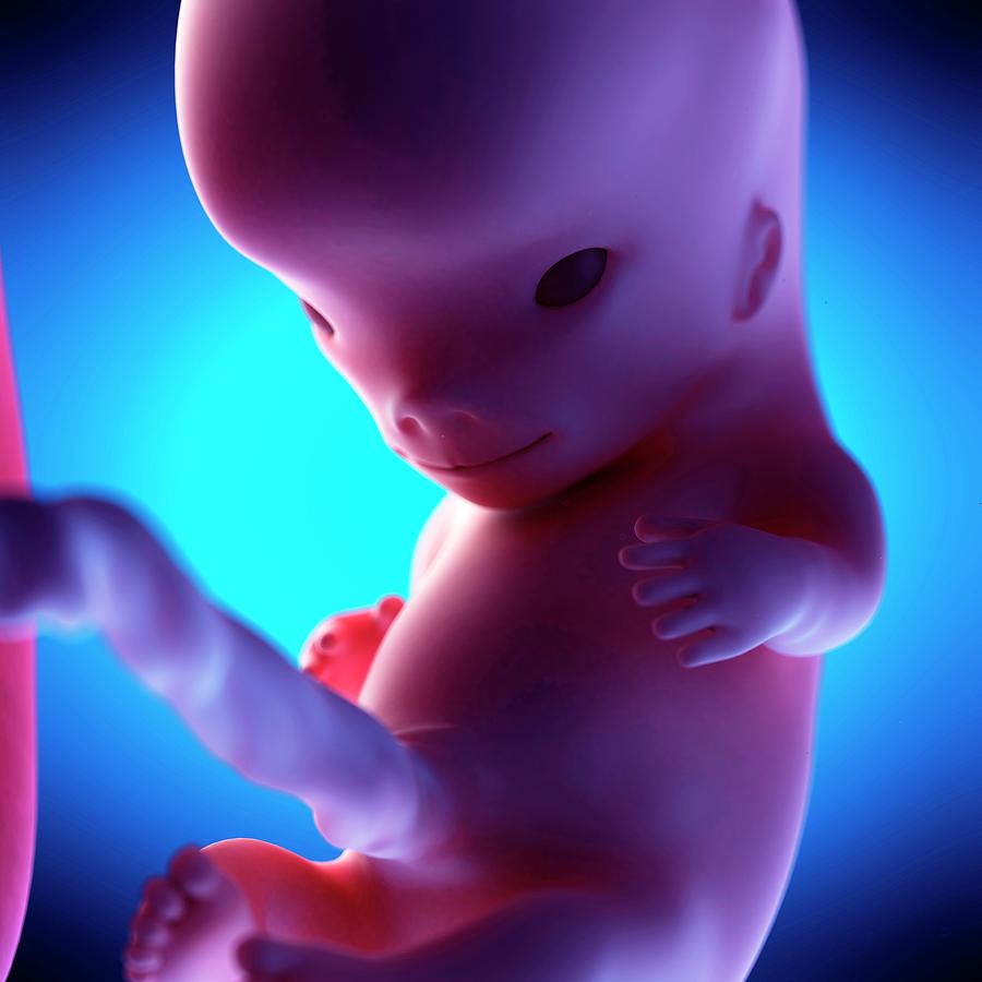
Human Fetus At Week 10 Of Gestation Photograph by Sebastian Kaulitzki
Fetus in fetu is a rare congenital anomaly and is defined as a monozygotic twin incorporated into the abdomen of its sibling during development. Fetus in fetu is often overlooked in the differential diagnosis of an abdominal mass. Unlike teratomas, fetus in fetu is a benign disorder. We describe the clinical characteristics of two patients, a thirty-months old boy who was found to have.
/168415756-56a771963df78cf77295fef3.jpg)
Early Pregnancy Fetal Development
Fetus-in-fetu: imaging and pathologic findings Abdom Imaging. 2012 Feb;37(1):147-50. doi: 10.1007/s00261-011-9757-2.. After opening the sac it was noted to contain an incompletely developed fetus with grossly visible limbs, clearly discernible male genitalia, hairs, and a poorly formed head. The fetus was connected to the sac via an 8 cm.
Labgrown embryos reveal earliest stages of life Literacy Project
Fetus-in-fetu. Imaging and pathology. 2012 Apr;33 (4):444-8. Fetus-in-fetu (FIF), also known as endoparasitic twin, is a form of asymmetric fetal duplication in which the abnormal developing embryo parasitizes the normal co-twin by attaching internally. Here, we report a case of FIF presented as an intra-abdominal cystic mass, which was first.

Fetus Pictures, Images and Stock Photos iStock
The term "fetus in fetu" was first described by Johann Friedrich Meckel in 1800 and defined by Willis in 1953 as a rare condition where a malformed parasitic twin resides in the body of its host. [ 2] However, there is controversy as to whether fetus in fetu is a distinct entity or represents a highly organized teratoma.

Normal Anatomy of the Fetus at MR Imaging RadioGraphics
Fetus in fetu is a rare congenital anomaly that occur secondary to abnormal embryogenesis in a diamniotic monochorionic pregnancy [1]. It was first described in the 1800s by Johann Friederich Meckle [2]. Less than 200 cases of fetus in fetu have been reported till now [3]. It is unusual condition in which a vertebrate fetus is enclosed within.
